

The upper airway in the era of GLP-1 receptor agonists: implications for obstructive sleep apnea in obesity
La vía aérea superior en la era de los agonistas del receptor del GLP-1: implicaciones para el síndrome de apnea obstructiva del sueño en la obesidad
[Artículo en inglés/ Article in English]
Vol. 10 Núm. 4. Agosto 2026 - Octubre 2026.
e-ISSN: 2530-5468 - Open Access Journal
DOI: 10.5281/zenodo.22965164
Published under Creative Commons CC BY-NC-ND 4.0


Sanum. vol. 10, número 4 (2026) páginas 92 – 103
AUTHORS:
Lucía Fernanda Vergara Pérez1 (0009-0009-7565-8097)
Jesús David Pérez Pabón1 (0009-0001-7826-2356)
Hilder Adrián Benavides Villamarín2 (0000-0002-3768-6304)
Valeria Ceballos García3 (0009-0002-8423-3225)
Adriana Lucía Calderón Silva4 (0009-0009-2348-6190)
María Paula Cabas Malaver5 (0009-0007-2914-4277)
Affiliations
- Facultad de Ciencias de la Salud, Universidad del Norte, Barranquilla, Colombia.
- Facultad de Ciencias de la Salud, Universidad del Cauca. Popayan, Colombia.
- Facultad de Ciencias de la Salud, Pontificia Universidad Javeriana, Cali, Colombia.
- Facultad de Medicina, Universidad Antonio Nariño, Bogotá, Colombia.
- Facultad de Medicina y Ciencias de la Salud, Universidad Militar Nueva Granada, Bogotá, Colombia.
Como citar este artículo:
Vergara Pérez LF, Pérez Pabón JD, Benavides Villamarín HA, Ceballos García V, Calderón Silva AL, Cabas Malaver MP. La vía aérea superior en la era de los agonistas del receptor del GLP-1: implicaciones para el síndrome de apnea obstructiva del sueño en la obesidad. SANUM 2026, 10(4) pp 92 – 103. DOI: 10.5281/zenodo.22965164
© Los autores. Publicado por SANUM: Revista Científico-Sanitaria bajo una licencia Creative Commons Atribución-NoComercial-SinDerivadas 4.0 Internacional (CC BY-NC-ND 4.0). 
How to cite this article:
Vergara Pérez LF, Pérez Pabón JD, Benavides Villamarín HA, Ceballos García V, Calderón Silva AL, Cabas Malaver MP. The upper airway in the era of GLP-1 receptor agonists: implications for obstructive sleep apnea in obesity. SANUM 2026, 10(4) pp 92 – 103. DOI: 10.5281/zenodo.22965164
© The authors. Published by SANUM: Revista Científico-Sanitaria under a Creative Commons Attribution-NonCommercial-NoDerivatives 4.0 International License (CC BY-NC-ND 4.0).
Article type: Review article.
Section: Pulmonology.
Received: 29-07-2026.
Accepted: 07-09-2026.
Published: 30-10-2026.
ABSTRACT
Introduction: Glucagon-like peptide-1 receptor agonists (GLP-1RAs) have been shown to significantly reduce the severity of obstructive sleep apnea (OSA). However, their potential effects on upper airway anatomy and physiology remain poorly understood. This review examines the available evidence regarding the role of GLP-1RAs in upper airway remodeling.
Methods: A narrative review was conducted using PubMed/MEDLINE, Scopus, and Google Scholar. Systematic reviews, meta-analyses, clinical trials, clinical practice guidelines, and experimental studies published between January 2000 and June 2026 were included.
Results: Obesity induces anatomical remodeling of the upper airway that promotes pharyngeal collapse. Evidence from bariatric surgery demonstrates that these changes are partially reversible and that tongue fat reduction is an important mediator of OSA improvement. GLP-1RAs consistently reduce visceral and ectopic adiposity and, in a pilot study, decreased tongue fat. However, direct evidence that these agents induce upper airway remodeling remains lacking.
Conclusions: GLP-1RAs may improve OSA not only through weight loss but also by promoting upper airway remodeling. Longitudinal studies incorporating imaging techniques and physiological assessments are needed to confirm this hypothesis.
KEYWORDS:
Glucagon-Like Peptide-1 Receptor Agonists;
Obesity;
Sleep Apnea, Obstructive;
Airway Management;
Bariatric Surgery.
RESUMEN
Introducción: Los agonistas del receptor del péptido similar al glucagón tipo 1 (GLP-1RA) han demostrado reducir significativamente la gravedad del síndrome de apnea obstructiva del sueño (SAHOS). Sin embargo, sus posibles efectos sobre la anatomía y fisiología de la vía aérea superior permanecen poco explorados. Esta revisión analiza la evidencia disponible sobre el papel de los GLP-1RA en el remodelado de la vía aérea superior.
Métodos: Se realizó una revisión narrativa mediante búsquedas en PubMed/MEDLINE, Scopus y Google Scholar. Se incluyeron revisiones sistemáticas, metaanálisis, ensayos clínicos, guías de práctica clínica y estudios experimentales publicados entre enero del 2000 y junio del 2026.
Resultados: La obesidad induce un remodelado anatómico de la vía aérea superior que favorece el colapso faríngeo. La evidencia derivada de la cirugía bariátrica demuestra que estos cambios son parcialmente reversibles y que la reducción de la grasa lingual constituye un mediador importante de la mejoría del SAHOS. Los GLP-1RA reducen la adiposidad visceral y ectópica y, en un estudio piloto, disminuyeron la grasa lingual; sin embargo, aún no existe evidencia directa de que induzcan un remodelado anatómico de la vía aérea superior.
Conclusiones: Los GLP-1RA podrían mejorar el SAHOS no solo mediante la pérdida de peso, sino también a través de un posible remodelado de la vía aérea superior. Estudios con técnicas de imagen y evaluaciones fisiológicas son necesarios para confirmar esta hipótesis.
PALABRAS CLAVE:
Agonistas del Receptor del GLP-1;
Obesidad;
Síndrome de Apnea Obstructiva del Sueño;
Manejo de la Vía Aérea;
Cirugía Bariátrica.
ABSTRACT
Introduction: Glucagon-like peptide-1 receptor agonists (GLP-1RAs) have been shown to significantly reduce the severity of obstructive sleep apnea (OSA). However, their potential effects on upper airway anatomy and physiology remain poorly understood. This review examines the available evidence regarding the role of GLP-1RAs in upper airway remodeling.
Methods: A narrative review was conducted using PubMed/MEDLINE, Scopus, and Google Scholar. Systematic reviews, meta-analyses, clinical trials, clinical practice guidelines, and experimental studies published between January 2000 and June 2026 were included.
Results: Obesity induces anatomical remodeling of the upper airway that promotes pharyngeal collapse. Evidence from bariatric surgery demonstrates that these changes are partially reversible and that tongue fat reduction is an important mediator of OSA improvement. GLP-1RAs consistently reduce visceral and ectopic adiposity and, in a pilot study, decreased tongue fat. However, direct evidence that these agents induce upper airway remodeling remains lacking.
Conclusions: GLP-1RAs may improve OSA not only through weight loss but also by promoting upper airway remodeling. Longitudinal studies incorporating imaging techniques and physiological assessments are needed to confirm this hypothesis.
KEYWORDS:
Glucagon-Like Peptide-1 Receptor Agonists;
Obesity;
Sleep Apnea, Obstructive;
Airway Management;
Bariatric Surgery.
INTRODUCTION:
Obstructive sleep apnea (OSA) is one of the most prevalent sleep-related breathing disorders worldwide. It is estimated that approximately one billion adults have some degree of OSA, with a prevalence ranging from 9% to 38% in the adult population, depending on the characteristics of the population studied and the diagnostic criteria applied (1,2). Its growing global burden has been closely linked to the increasing prevalence of obesity and is associated with substantial health, social, and economic consequences (3). In the United States alone, the annual cost attributable to untreated OSA is estimated to exceed 150 billion US dollars, underscoring the magnitude of this disease and the need for more effective therapeutic strategies (1).
Obesity is the leading modifiable risk factor for the development of OSA and has a bidirectional relationship with the disease, as it promotes OSA development while OSA itself may contribute to further weight gain through metabolic and behavioral alterations (4,5). The risk of OSA increases progressively with body mass index (BMI); even a 10% increase in body weight has been associated with a 32% increase in the apnea-hypopnea index (AHI) (6), whereas individuals with obesity have a four- to tenfold higher risk of developing OSA compared with those of normal weight (3,7,8). Consequently, nearly 70% of individuals with obesity have some degree of OSA (9,10). Nevertheless, obesity does not account for all cases, as a recent meta-analysis showed that approximately one quarter of patients with OSA have normal body weight and nearly 70% do not meet criteria for obesity (3).
Traditionally, obesity management has relied on intensive lifestyle interventions. Although the combination of dietary modification, physical activity, and behavioral therapy achieves weight reductions of approximately 8% of total body weight (11), long-term weight maintenance remains challenging, with progressive weight regain typically beginning around 36 weeks after completion of the intervention (12). Bariatric surgery has therefore represented the most effective treatment for sustained weight loss, achieving total body weight reductions ranging from approximately 19% to 26% at five years, depending on the surgical procedure (approximately 25.5% following Roux-en-Y gastric bypass, 18.8% after sleeve gastrectomy, and 11.7% after adjustable gastric banding) (13). However, its invasive nature, perioperative risks, limited access, lack of social support, short- and long-term gastrointestinal complications, and low patient acceptance have resulted in fewer than 1% of eligible individuals undergoing bariatric surgery each year (14–16). The introduction of glucagon-like peptide-1 receptor agonists (GLP-1RAs), and more recently dual glucose-dependent insulinotropic polypeptide (GIP)/GLP-1 receptor agonists, has transformed the pharmacological management of obesity by achieving weight reductions of 15–20% (17), outcomes that were previously attainable almost exclusively through bariatric surgery.
Interest in this field has intensified following the publication of the SURMOUNT-OSA trials, which demonstrated that tirzepatide significantly reduces the apnea-hypopnea index in patients with obesity and moderate-to-severe OSA, providing the first robust evidence that a pharmacological therapy can effectively treat this disorder (18). However, most available reviews have focused on the magnitude of weight loss and the reduction in the apnea-hypopnea index, interpreting these findings primarily as a consequence of overall weight reduction. In contrast, considerably less attention has been paid to the potential effects of these therapies on upper airway anatomy and physiology, and to the implications that these changes may have for contemporary OSA management. In this review, we synthesize the available evidence from an upper airway–centered perspective and discuss how GLP-1 receptor agonists may reshape our understanding of OSA pathophysiology and influence future therapeutic strategies.
METHODS
A narrative review of the scientific literature was conducted to evaluate the impact of glucagon-like peptide-1 receptor agonists (GLP-1RAs) on obstructive sleep apnea (OSA), with particular emphasis on upper airway–related pathophysiological mechanisms and their clinical implications. A literature search was performed in the PubMed/MEDLINE, Scopus, and Google Scholar databases, including publications from January 2000 through June 2026. Combinations of Medical Subject Headings (MeSH) and Health Sciences Descriptors (DeCS) were used, including the terms obstructive sleep apnea, sleep-disordered breathing, glucagon-like peptide-1 receptor agonists, GLP-1 receptor agonists, semaglutide, tirzepatide, obesity, upper airway, pharyngeal collapsibility, weight loss, and upper airway anatomy, as well as their Spanish equivalents, using the Boolean operators AND and OR. Studies were included if they addressed the effects of GLP-1RAs on OSA, obesity-related upper airway anatomy or physiology, or mechanisms relevant to upper airway remodeling. Publications unrelated to the review objectives, non-peer-reviewed reports, conference abstracts without full-text availability, and duplicate records were excluded. Priority was given to systematic reviews, meta-analyses, randomized controlled trials, clinical practice guidelines, consensus statements, and relevant experimental or physiological studies based on their methodological quality and relevance to the objectives of this review.
RESULTS
- The upper airway in obesity.
1.1. Obesity-Induced anatomical remodeling of the upper airway
Obesity induces profound anatomical remodeling of the upper airway, providing the principal structural substrate for the development of obstructive sleep apnea (OSA). Increased adiposity promotes ectopic fat deposition within and around the pharynx, resulting in enlargement of multiple soft-tissue structures, including the tongue, soft palate, uvula, lateral pharyngeal walls, and parapharyngeal fat pads (19).
Among these alterations, tongue fat accumulation represents one of the most important anatomical changes. Fat deposition within the tongue, particularly in the genioglossus muscle, increases tongue volume and promotes posterior displacement of the tongue base as muscle tone decreases during sleep, thereby reducing the retrolingual airway space (20). In addition, increased tongue volume has been associated with inferior displacement of the hyoid bone, further contributing to narrowing of the pharyngeal lumen (21). Likewise, fat accumulation within the parapharyngeal fat pads exerts mechanical pressure on the lateral pharyngeal walls, promoting upper airway narrowing and acting as an independent determinant of OSA severity, particularly in patients with a predominant pattern of lateral pharyngeal collapse (22).
Obesity also alters the morphology of the soft palate and lateral pharyngeal walls. Enlargement of these structures, associated with fatty infiltration and elongation of the soft palate, reduces the retropalatal airway space, whereas thickening of the lateral pharyngeal walls further increases the mechanical load imposed on the upper airway (21). More recently, three-dimensional magnetic resonance imaging studies have demonstrated that increasing body mass index is accompanied by a progressive enlargement of virtually all pharyngeal soft tissues, including the tongue, soft palate, lateral pharyngeal walls, parapharyngeal fat pads, epiglottis, and pterygoid muscles. These structural changes are accompanied by a characteristic pattern of upper airway narrowing, with a predominant reduction in the lateral airway diameter, particularly at the retropalatal level (23).
From a clinical perspective, neck circumference serves as an indirect marker of this anatomical remodeling because it reflects adipose tissue accumulation surrounding the upper airway and is strongly associated with both the risk and severity of OSA (24,25). As a consequence of these structural alterations, the upper airway undergoes a progressive reduction in luminal caliber and anatomical reserve against collapse. Importantly, this narrowing is not solely attributable to local fat deposition but also to indirect obesity-related mechanisms, including reductions in lung volumes caused by central adiposity, which diminish the caudal traction exerted on the pharynx (21,23). Collectively, these changes establish the anatomical substrate upon which the functional abnormalities responsible for upper airway collapse during sleep develop.
1.2. Functional Consequences of Upper Airway Remodeling
The anatomical alterations described above translate into functional changes that increase the susceptibility of the pharynx to collapse during sleep (4). Unlike other segments of the respiratory tract, the pharynx lacks rigid bony or cartilaginous support and behaves as a collapsible tube whose stability depends on the interaction among intraluminal pressure, the mechanical properties of its surrounding tissues, and the activity of the upper airway dilator muscles. Consequently, obesity-induced anatomical remodeling increases the mechanical load imposed on the upper airway and promotes greater pharyngeal collapsibility, which is considered one of the principal pathophysiological mechanisms underlying OSA (26).
The most objective measure of this propensity for collapse is the pharyngeal critical closing pressure (Pcrit), defined as the intraluminal pressure at which the upper airway completely collapses. Pcrit is regarded as the principal physiological marker of pharyngeal collapsibility because it integrates the combined effects of multiple anatomical determinants, including peripharyngeal adiposity, tongue volume, pharyngeal length, hyoid bone position, and upper airway dimensions (27,28). In patients with obesity and OSA, more positive Pcrit values indicate a greater tendency toward upper airway collapse and correlate with both disease severity and anthropometric measures such as body mass index and neck circumference (28).
Upper airway stability also depends on dynamic factors that modulate the mechanical forces acting on the pharynx. Among these, lung volume plays a pivotal role. Central adiposity reduces functional residual capacity and expiratory reserve volume as a consequence of decreased chest wall compliance and cephalad displacement of the diaphragm, effects that become more pronounced in the supine position and during sleep (29). As a result, the caudal traction exerted by the trachea on the pharynx is diminished, reducing longitudinal tension within the pharyngeal walls and increasing their susceptibility to collapse (30). This mechanism is particularly relevant in individuals with severe obesity, in whom the reduction in lung volume is more pronounced and contributes substantially to increased pharyngeal collapsibility.
To counterbalance these mechanical loads, compensatory mechanisms are activated to preserve upper airway patency through increased activity of the pharyngeal dilator muscles. During wakefulness, reflex activation of the genioglossus and other upper airway dilator muscles partially compensates for anatomical narrowing and helps maintain pharyngeal patency. However, this compensatory response declines during sleep onset and throughout sleep progression as a consequence of the physiological reduction in neural drive to the dilator muscles and decreased responsiveness to negative intrapharyngeal pressure, allowing collapsing forces to predominate (31). Beyond this functional impairment of neuromuscular compensation, emerging evidence suggests that obesity may also induce structural changes within the genioglossus muscle, including intramuscular lipid infiltration, remodeling of muscle fiber composition, and alterations in muscle architecture. These changes may reduce fatigue resistance and impair the muscle’s ability to respond to the increased mechanical load imposed during sleep (31,32). Collectively, these findings support the concept that obesity compromises upper airway stability not only through anatomical remodeling but also by impairing the neuromuscular mechanisms responsible for maintaining airway patency. The anatomical and physiological changes affecting the upper airway in obesity are summarized in Figure 1.
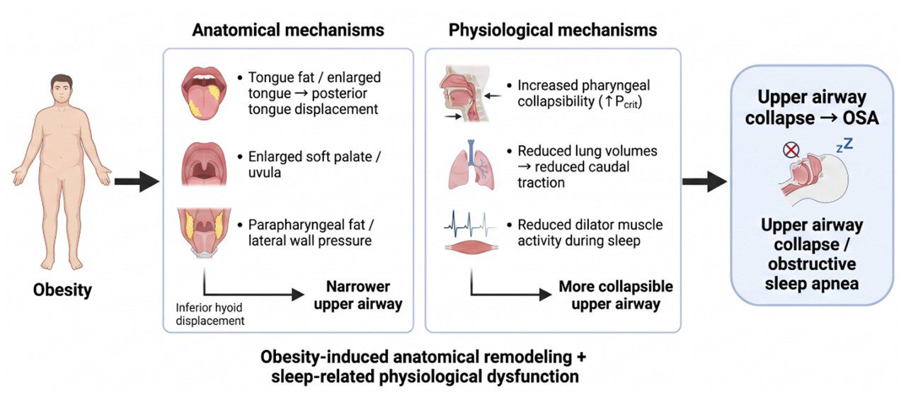
Figure 1. Anatomical and physiological mechanisms linking obesity to upper airway collapse. Source: Developed by the authors. Created with BioRender.com (https://biorender.com/shortURL).
1.3. Obesity as a Modifiable Anatomical Trait
Weight loss is the cornerstone of reversing obesity-induced upper airway remodeling. Evidence from lifestyle interventions and bariatric surgery has consistently demonstrated that weight reduction induces measurable anatomical remodeling of the upper airway, characterized by reductions in soft-tissue volume, decreased ectopic fat deposition, enlargement of the pharyngeal airway, and reduced upper airway collapsibility. These structural changes are accompanied by significant improvements in the severity of obstructive sleep apnea (OSA) (33–35).
Imaging studies have provided detailed characterization of this reverse remodeling process. Following weight loss, reductions have been observed in the volume of multiple upper airway soft tissues, including the tongue, soft palate, lateral pharyngeal walls, pterygoid muscles, and parapharyngeal fat pads, together with enlargement of the velopharyngeal space and increased retropalatal airway caliber. Beyond reductions in tissue volume, magnetic resonance imaging with fat quantification has demonstrated decreased intratissue fat infiltration within the tongue, tongue base, lateral pharyngeal walls, and soft palate, indicating that weight loss modifies not only tissue size but also tissue composition (33,34,36).
A particularly important finding is that improvement in OSA is not solely determined by the magnitude of weight loss. Several studies have shown that reductions in ectopic fat within the upper airway, particularly tongue fat, account for a substantial proportion of the improvement in the apnea-hypopnea index (34). Notably, reductions in tongue fat volume remain significantly associated with clinical improvement even after adjustment for the total amount of weight lost, suggesting that regional fat redistribution may be a more important determinant of OSA improvement than overall weight reduction (34,37). These findings support the hypothesis that tongue fat represents one of the principal anatomical mediators linking obesity to pharyngeal collapse.
The anatomical remodeling induced by weight loss also has important functional consequences. Reduction in the volume of pharyngeal soft tissues decreases the mechanical loads that promote upper airway collapse, increases airway caliber, and reduces pharyngeal collapsibility, changes that are reflected by a reduction in pharyngeal critical closing pressure (Pcrit) (37). Although the magnitude of this response varies among individuals and depends on its interaction with other pathophysiological traits, the available evidence supports the concept that the anatomical component of OSA is dynamic and potentially reversible.
- GLP-1 receptor agonists: beyond weight loss
2.1. Effects on Body Composition
Glucagon-like peptide-1 receptor agonists (GLP-1RAs) induce substantial weight loss accompanied by favorable changes in body composition that extend beyond reductions in total body weight. Multiple randomized clinical trials and meta-analyses have demonstrated that most of the weight loss achieved with these agents is attributable to reductions in fat mass, with relative preservation of lean mass (38,39). In a recent meta-analysis, GLP-1RAs produced an approximate 9% reduction in body weight after three months of treatment, accompanied by significant decreases in total fat mass and visceral adipose tissue, whereas reductions in lean mass were considerably smaller (39). Similarly, in the SURMOUNT-1 trial, approximately 75% of the weight loss achieved with tirzepatide consisted of fat mass, whereas the remaining 25% corresponded to lean mass, a distribution that was virtually identical to that observed with placebo. These findings suggest that these agents promote favorable body composition remodeling without disproportionately increasing lean tissue loss (40). Consistent with these observations, GLP-1RAs also reduce waist circumference, reflecting a preferential reduction in abdominal adiposity (39).
Beyond reducing total fat mass, robust evidence indicates that GLP-1RAs significantly decrease visceral adipose tissue, one of the most metabolically active fat depots and a key contributor to insulin resistance, systemic inflammation, and cardiometabolic risk. A meta-analysis of 30 randomized controlled trials involving 1,736 participants demonstrated a significant reduction in visceral adipose tissue compared with control groups (standardized mean difference [SMD] = −0.59; 95% confidence interval [CI], −0.83 to −0.36). This effect was consistently observed in patients with type 2 diabetes mellitus (SMD = −0.49), metabolic dysfunction-associated steatotic liver disease (SMD = −0.99), and even in individuals without diabetes (SMD = −1.38) (41). Consistent with these findings, the body composition substudy of the SURMOUNT-1 trial demonstrated significantly greater reductions in visceral fat mass with tirzepatide than with placebo after 72 weeks of treatment (42).
In addition to visceral adiposity, GLP-1RAs reduce several ectopic fat depots, particularly hepatic fat content. A meta-analysis demonstrated a significant reduction in liver fat content compared with control groups (weighted mean difference [WMD] = −3.09; 95% CI, −4.16 to −2.02) (41), while studies evaluating semaglutide and liraglutide confirmed sustained reductions in hepatic steatosis assessed by imaging techniques, accompanied by improvements in metabolic parameters (43,44). From the perspective of obstructive sleep apnea (OSA), these findings are particularly relevant because they demonstrate that GLP-1RAs consistently modify regional fat distribution and reduce ectopic fat depots beyond subcutaneous adipose tissue.
2.2. Could GLP-1 Receptor Agonists Induce Upper Airway Remodeling?
The demonstration that weight loss induced by bariatric surgery reverses anatomical remodeling of the upper airway (33–35) raises an important pathophysiological question: can glucagon-like peptide-1 receptor agonists (GLP-1RAs) induce similar structural changes? Although the available evidence remains limited, current findings suggest that this possibility is biologically plausible and warrants further investigation.
Until recently, this hypothesis was supported exclusively by indirect evidence derived from reductions in visceral and ectopic adiposity, together with the clinical improvement observed in patients with obstructive sleep apnea (OSA) (41). In a randomized, two-arm pilot trial involving 25 women with obesity and polycystic ovary syndrome, 16 weeks of treatment with semaglutide produced a significant reduction in tongue fat volume (−1.94 ± 5.51 cm³ vs. +3.12 ± 4.87 cm³ with placebo; p = 0.022) and tongue fat fraction (−0.02 ± 0.07 vs. +0.04 ± 0.06; p = 0.010). These changes correlated with reductions in body weight, body mass index, and waist circumference (45). Interestingly, total tongue volume did not change significantly, suggesting that semaglutide primarily reduced fat content rather than overall tongue size. The authors concluded that these findings provide the first evidence that a GLP-1 receptor agonist can directly modify tongue structure, while acknowledging the exploratory nature of the study and the need for larger confirmatory investigations.
Beyond this pilot study, the available evidence remains predominantly indirect. The SURMOUNT-OSA trials demonstrated marked reductions in OSA severity, including decreases in the apnea-hypopnea index of approximately 51–59%, absolute reductions of 20–24 events/hour compared with placebo, weight loss of 17.7–19.6%, and substantial reductions in hypoxic burden (18). Consistent with these findings, recent meta-analyses have reported mean reductions in the apnea-hypopnea index ranging from 13.9 to 14.5 events/hour with GLP-1 receptor agonists compared with control groups (46,47). However, none of these studies incorporated magnetic resonance imaging, computed tomography, or other imaging modalities specifically designed to directly assess anatomical changes in the upper airway.
The available evidence supports a compelling yet still incomplete pathophysiological hypothesis. GLP-1 receptor agonists consistently reduce visceral adiposity, several ectopic fat depots, and, in at least one pilot study, tongue fat, one of the principal anatomical determinants of OSA. However, it remains unknown whether these changes translate into reductions in parapharyngeal fat, enlargement of the upper airway, reductions in pharyngeal critical closing pressure (Pcrit), or modifications of other anatomical traits associated with pharyngeal collapse. The lack of longitudinal studies incorporating magnetic resonance imaging or computed tomography, as well as mediation analyses comparable to those performed after bariatric surgery, represents one of the major knowledge gaps in this field and should be considered a priority for future research. The Table I summarizes the current evidence supporting upper airway remodeling following bariatric surgery and GLP-1 receptor agonist therapy.
| Remodeling feature | Evidence after bariatric surgery | Evidence after GLP-1 receptor agonists |
| Magnitude of weight loss | Consistent and sustained (20–30% total body weight loss) | Consistent (15–20% total body weight loss with semaglutide/tirzepatide) |
| Reduction in tongue fat | Demonstrated by MRI; independently associated with improvement in AHI | Preliminary evidence from one MRI pilot study showing reduced tongue fat volume and fat fraction |
| Reduction in parapharyngeal fat | Demonstrated by MRI | Not evaluated |
| Reduction in soft palate volume | Demonstrated | Not evaluated |
| Reduction in lateral pharyngeal wall thickness | Demonstrated | Not evaluated |
| Increase in upper airway caliber | Demonstrated by MRI/CT | Not evaluated |
| Reduction in pharyngeal collapsibility (Pcrit) | Demonstrated in physiological studies | Not evaluated |
| Improvement in upper airway anatomy by imaging | Multiple MRI and CT studies | Limited to a single exploratory MRI study |
| Mediation analysis linking anatomical remodeling with AHI improvement | Demonstrated (tongue fat identified as a major mediator) | Not available |
| Direct evidence of upper airway remodeling | Strong | Limited |
| Overall level of mechanistic evidence | High | Low to moderate |
Table I. Current evidence supporting upper airway remodeling following bariatric surgery and GLP-1 receptor agonist therapy. MRI, magnetic resonance imaging; CT, computed tomography; Pcrit, pharyngeal critical closing pressure; AHI, apnea-hypopnea index. Source: Created by the authors
2.3. Additional Mechanisms Potentially Contributing to OSA Improvement
Although weight loss and the potential anatomical remodeling of the upper airway represent the most apparent mechanisms underlying the improvement of obstructive sleep apnea (OSA) with glucagon-like peptide-1 receptor agonists (GLP-1RAs), accumulating evidence indicates that these agents exert additional systemic effects that may also contribute to their clinical benefits. Among these, their anti-inflammatory effects are the best documented. Recent meta-analyses have demonstrated significant reductions in C-reactive protein (standardized mean difference [SMD] = −0.37; 95% confidence interval [CI], −0.59 to −0.14), tumor necrosis factor-α, and interleukin-6, accompanied by an increase in adiponectin (SMD = 0.30; 95% CI, 0.13–0.46) (48,49). Consistent with these findings, the SURMOUNT-OSA trials reported reductions of 0.7–1.0 mg/L in high-sensitivity C-reactive protein compared with placebo, together with decreases in systolic blood pressure, supporting anti-inflammatory and vasculoprotective effects that extend beyond weight loss alone (18,48,50). In addition, GLP-1 receptor agonists reduce markers of oxidative stress, particularly malondialdehyde (SMD = −0.98; 95% CI, −1.65 to −0.30), which may attenuate the endothelial injury induced by the intermittent hypoxia characteristic of OSA (48). It has also been proposed that reductions in visceral adiposity may improve respiratory mechanics by decreasing diaphragmatic restriction and increasing lung volumes. However, this hypothesis is based primarily on indirect evidence, and no studies have yet demonstrated improvements in physiological parameters such as functional residual capacity, expiratory reserve volume, or pharyngeal critical closing pressure (Pcrit) (51,52).
Several physiological mechanisms implicated in the pathogenesis of OSA remain largely unexplored in the context of GLP-1 receptor agonist therapy. Although these agents exert well-established central effects through hypothalamic and brainstem circuits involved in appetite regulation and energy homeostasis (53,54), it remains unknown whether these neuronal actions also influence ventilatory control, chemosensitivity, loop gain, or arousal threshold during sleep. Likewise, no studies have evaluated the effects of GLP-1 receptor agonists on nocturnal rostral fluid shift or on objective sleep architecture assessed by polysomnography. Consequently, any contribution of these mechanisms to the improvement in OSA observed with GLP-1 receptor agonists remains speculative.
DISCUSSION
Glucagon-like peptide-1 receptor agonists (GLP-1RAs) have transformed the pharmacological management of obesity (15) and, more recently, have demonstrated significant clinical efficacy in patients with obstructive sleep apnea (OSA) (47). For decades, weight loss was regarded as a nonspecific determinant of OSA improvement. However, imaging studies conducted after bariatric surgery have shown that reductions in ectopic fat within the upper airway, particularly tongue fat, represent a key anatomical mediator of this improvement (33,34,36). Emerging evidence suggests that GLP-1 receptor agonists may induce a similar process of anatomical remodeling, shifting the focus from overall weight loss toward specific structural changes within the upper airway (45).
Despite this biological plausibility, the available evidence remains limited. To date, only a single pilot study has demonstrated reductions in tongue fat assessed by magnetic resonance imaging following semaglutide treatment (45), whereas large clinical trials and published meta-analyses have focused primarily on clinical outcomes such as the apnea-hypopnea index, hypoxic burden, and weight loss (18). Consequently, it remains unclear to what extent the improvement in OSA is attributable to specific anatomical changes in the upper airway, global reductions in adiposity, or other physiological mechanisms associated with treatment. This represents an important gap between the demonstration of clinical efficacy and the understanding of the mechanisms responsible for these therapeutic benefits.
Several fundamental pathophysiological traits of OSA remain largely unexplored in the context of GLP-1 receptor agonist therapy. Although these agents exert well-established anti-inflammatory and antioxidant effects (48,49) and may indirectly improve respiratory mechanics through reductions in central adiposity (46,47), no studies have systematically evaluated their impact on key physiological parameters such as pharyngeal collapsibility, pharyngeal critical closing pressure (Pcrit), loop gain, arousal threshold, or the compensatory responses of the upper airway dilator muscles. The absence of these data limits the ability to identify which patient phenotypes are most likely to benefit from these therapies and hinders progress toward a truly personalized approach to OSA management.
If future studies confirm that GLP-1 receptor agonists induce meaningful anatomical remodeling of the upper airway, several aspects of contemporary OSA management may need to be reconsidered. Reassessment of disease severity following substantial weight loss, the need for repeat sleep studies, adjustments in continuous positive airway pressure (CPAP) requirements, and the selection of candidates for upper airway-targeted therapies may all become increasingly relevant. Rather than replacing established treatments, GLP-1 receptor agonists are likely to become an additional component of a multidisciplinary, phenotype-based approach to OSA management.
Future Directions
Although the clinical benefits of GLP-1 receptor agonists in patients with obesity and OSA have now been demonstrated, important pathophysiological and clinical questions remain unanswered. Key priorities for future research include:
- What degree of weight loss is required to induce clinically meaningful upper airway remodeling?
- Does reduction in tongue fat account for a substantial proportion of the improvement in OSA observed with GLP-1 receptor agonist therapy?
- Do these agents modify other anatomical structures involved in upper airway collapse, such as the parapharyngeal fat pads, soft palate volume, or pharyngeal airway caliber?
- Do GLP-1 receptor agonists influence non-anatomical pathophysiological traits, including loop gain, arousal threshold, or the neuromuscular responsiveness of the upper airway?
- Should magnetic resonance imaging, computed tomography, or drug-induced sleep endoscopy (DISE) be incorporated as outcome measures in future clinical trials?
- Could reductions in tongue fat serve as a biomarker of treatment response?
- Which patients will continue to require upper airway-targeted therapies despite achieving substantial weight loss with GLP-1 receptor agonists?
CONCLUSIONS
The available evidence suggests that glucagon-like peptide-1 receptor agonists (GLP-1RAs) represent far more than a weight-loss strategy for patients with obesity and obstructive sleep apnea (OSA). Current data support the hypothesis that these agents may promote anatomical remodeling of the upper airway through reductions in ectopic fat deposition, particularly tongue fat. However, direct evidence remains limited, and important knowledge gaps persist regarding their effects on other anatomical and physiological components of the upper airway. Future studies incorporating advanced imaging techniques and comprehensive physiological assessments will be essential to confirm this hypothesis and to define the role of GLP-1 receptor agonists within a precision medicine approach to OSA.
DECLARATIONS:
Funding
This work received no funding.
Conflicts of interest
The authors declare that they have no conflicts of interest related to the content of this article.
Data availability
As this is a review article based on published scientific literature, no original datasets were generated. The information used derives entirely from the bibliographic sources cited in the manuscript.
Author contributions (CRediT)
LFVP: Conceptualization, Methodology, Writing – original draft, Writing – review & editing, Supervision, Visualization.
JDPP: Conceptualization, Methodology, Writing – original draft, Writing – review & editing, Supervision, Visualization.
HABV: Conceptualization, Methodology, Writing – review & editing, Visualization.
VCG: Conceptualization, Methodology, Writing – review & editing, Visualization.
ALCS: Conceptualization, Methodology, Writing – review & editing, Visualization.
MPCM: Conceptualization, Methodology, Writing – review & editing, Visualization.
- LFVP: Lucía Fernanda Vergara Pérez.
- JDPP: Jesús David Pérez Pabón.
- HABV: Hilder Adrián Benavides Villamarín.
- VCG: Valeria Ceballos García.
- ALCS: Adriana Lucía Calderón Silva.
- MPCM: María Paula Cabas Malaver.
The authors participated in the preparation of the manuscript according to the roles described by the CRediT taxonomy, critically reviewed the manuscript, approved the final version for publication, and accept responsibility for all aspects of the work in accordance with the criteria established by the International Committee of Medical Journal Editors (ICMJE).
Use of Artificial Intelligence
The authors declare that no artificial intelligence tools were used in the preparation of this manuscript.
Previous publication
This work is original and unpublished and has not been previously published or submitted simultaneously to another journal for publication. It has not been previously presented at congresses, scientific meetings, or other scientific events.
Ethical considerations
As this is a narrative review based exclusively on published scientific literature and does not include primary data from humans or animals, evaluation by a Research Ethics Committee was not required.
Transparency statement
The authors declare that the information presented in the manuscript is complete and transparent and that the results of the review have been interpreted objectively. They also undertake to notify the journal of any errors or deficiencies that may be detected before or after publication.
REFERENCES:
- Iannella G, Pace A, Bellizzi MG, Magliulo G, Greco A, De Virgilio A, et al. The Global Burden of Obstructive Sleep Apnea. Diagnostics (Basel). 2025;15(9):1088. doi: 10.3390/diagnostics15091088.
- Benjafield A V., Ayas NT, Eastwood PR, Heinzer R, Ip MSM, Morrell MJ, et al. Estimation of the global prevalence and burden of obstructive sleep apnoea: a literature-based analysis. Lancet Respir Med. 2019;7(8):687–98. doi:10.1016/S2213-2600(19)30198-5.
- Esmaeili N, Gell L, Imler T, Hajipour M, Taranto-Montemurro L, Messineo L, et al. The relationship between obesity and obstructive sleep apnea in four community-based cohorts: an individual participant data meta-analysis of 12,860 adults. EClinicalMedicine. 2025;83:103221. doi: 10.1016/j.eclinm.2025.103221.
- Veasey SC, Rosen IM. Obstructive Sleep Apnea in Adults. New England Journal of Medicine. 2019;380(15):1442–9. doi:10.1056/nejmcp1816152.
- Pillar G, Shehadeh N. Abdominal fat and sleep apnea: the chicken or the egg? Diabetes Care. 2008;31 Suppl 2(7):S303-9. doi: 10.2337/dc08-s272.
- Peppard PE, Young T, Palta M, Dempsey J, Skatrud J. Longitudinal study of moderate weight change and sleep-disordered breathing. JAMA. 2000;284(23):3015-21. doi: 10.1001/jama.284.23.3015.
- Meyer EJ, Wittert GA. Approach the Patient With Obstructive Sleep Apnea and Obesity. Journal of Clinical Endocrinology and Metabolism. 2024;109(3):e1267–79. doi:10.1210/clinem/dgad572.
- Lyu YS, Lee JH, Yoon Y, Kim JH, Kim SY. Association Between Sleep Apnea Risk and Obesity Phenotypes in Korean Adults: A Nationwide Population-Based Study. J Clin Med. 2026;15(9). doi:10.3390/jcm15093240
- Jehan S, Zizi F, Pandi-Perumal SR, Wall S, Auguste E, Myers AK, Jean-Louis G, McFarlane SI. Obstructive Sleep Apnea and Obesity: Implications for Public Health. Sleep Med Disord. 2017;1(4):00019.
- Messineo L, Bakker JP, Cronin J, Yee J, White DP. Obstructive sleep apnea and obesity: A review of epidemiology, pathophysiology and the effect of weight-loss treatments. Sleep Med Rev. 2024;78:101996. doi: 10.1016/j.smrv.2024.101996.
- Webb VL, Wadden TA. Intensive Lifestyle Intervention for Obesity: Principles, Practices, and Results. Gastroenterology. 2017;152(7):1752–64. doi:10.1053/j.gastro.2017.01.045.
- Machado AM, Guimarães NS, Bocardi VB, da Silva TPR, Carmo AS do, Menezes MC de, et al. Understanding weight regain after a nutritional weight loss intervention: Systematic review and meta-analysis. Clin Nutr ESPEN. 2022;49:138–53. doi:10.1016/j.clnesp.2022.03.020.
- Arterburn D, Wellman R, Emiliano A, Smith SR, Odegaard AO, Murali S, et al. Comparative effectiveness and safety of bariatric procedures for weight loss a pcornet cohort study. Ann Intern Med. 2018;169(11):741–50. doi:10.7326/M17-2786.
- Murtha JA, Alagoz E, Breuer CR, Finn A, Raffa SD, Voils CI, et al. Individual-level barriers to bariatric surgery from patient and provider perspectives: A qualitative study. Am J Surg. 2022;224(1):429–36. doi:10.1016/j.amjsurg.2021.12.022.
- Dawwas GK, Samuels JM, Stein CM. Shifting obesity treatment paradigms: Trends of glucagon-like peptide-1 receptor agonists and bariatric surgery in the United States. Diabetes Obes Metab. 2025;27(10):6023-6026. doi:10.1111/dom.16600.
- Keogh N, Horsom D, Lee G, Saab MM. Factors that influence an individual’s decision to undergo bariatric surgery: A qualitative systematic review. PLoS One. 2025;20(10):e0334837. doi: 10.1371/journal.pone.0334837.
- Vienghirun J, Sawangjit R, Suttiruksa S, Chaiyakunapruk N, Jinathongthai P, Khunti K, et al. GLP-1 Receptor/Dual Agonists for Weight Loss: A Systematic Review and Network Meta-Analysis of RCTs. Diabetes Obes Metab. 2026;28(6):5043–57. doi:10.1111/dom.70698.
- Malhotra A, Grunstein RR, Fietze I, Weaver TE, Redline S, Azarbarzin A, et al. Tirzepatide for the Treatment of Obstructive Sleep Apnea and Obesity. New England Journal of Medicine. 2024;391(13):1193–205. doi:10.1056/nejmoa2404881
- Drager LF, Togeiro SM, Polotsky VY, Lorenzi-Filho G. Obstructive sleep apnea: a cardiometabolic risk in obesity and the metabolic syndrome. J Am Coll Cardiol. 2013;62(7):569-76. doi: 10.1016/j.jacc.2013.05.045.
- Kim AM, Keenan BT, Jackson N, Chan EL, Staley B, Poptani H, et al. Tongue fat and its relationship to obstructive sleep apnea. Sleep. 2014;37(10):1639-1648D. doi:10.5665/sleep.4072.
- Gottlieb DJ, Punjabi NM. Diagnosis and Management of Obstructive Sleep Apnea: A Review. JAMA – Journal of the American Medical Association. 2020;323(14):1380–400. doi:10.1001/jama.2020.3514
- Chen HC, Wang CJ, Lo YL, Hsu HC, Huang CG, Kuo IC, et al. Parapharyngeal fat pad area at the subglosso-supraglottic level is associated with corresponding lateral wall collapse and apnea-hypopnea index in patients with obstructive sleep apnea: a pilot study. Sci Rep. 2019;9(1):17722. doi: 10.1038/s41598-019-53515-5.
- Xu L, Keenan BT, Wiemken AS, Staley B, Benediktsdottir B, Juliusson S, et al. The Relationship between Upper Airway Anatomy and Obesity in Patients with Obstructive Sleep Apnea. Ann Am Thorac Soc. 2025;22(11):1749–63. doi:10.1513/AnnalsATS.202410-1027OC.
- Ibrahim MIS, Mohamad H, Mohamad A, Mohamad I. Association between neck circumference and the severity of obstructive sleep apnea. Polish Annals of Medicine. 2020;27(1):1–6. doi:10.29089/2020.20.00097
- Tom C, Roy B, Vig R, Kang DW, Aysola RS, Woo MA, et al. Correlations Between Waist and Neck Circumferences and Obstructive Sleep Apnea Characteristics. Sleep Vigil. 2018;2(2):111–8. doi:10.1007/s41782-018-0041-1
- Schwartz AR, Patil SP, Squier S, Schneider H, Kirkness JP, Smith PL, et al. Pulmonary Physiology and Pathophysiology of Obesity Obesity and upper airway control during sleep. J Appl Physiol. 2010;108:430–5. doi:10.1152/japplphysiol.00919.2009.
- Genta PR, Schorr F, Eckert DJ, Gebrim E, Kayamori F, Moriya HT, et al. Upper airway collapsibility is associated with obesity and hyoid position. Sleep. 2014;37(10):1673–8. doi:10.5665/sleep.4078.
- Kazemeini E, Van de Perck E, Dieltjens M, Willemen M, Verbraecken J, Op de Beeck S, et al. Critical to Know Pcrit: A Review on Pharyngeal Critical Closing Pressure in Obstructive Sleep Apnea. Front Neurol. 2022;13:775709. doi: 10.3389/fneur.2022.775709.
- Goligher EC, Ferguson ND, Brochard LJ. Clinical challenges in mechanical ventilation. Lancet. 2016;387(10030):1856-66. doi: 10.1016/S0140-6736(16)30176-3.
- Isono S. Obesity and obstructive sleep apnoea: mechanisms for increased collapsibility of the passive pharyngeal airway. Respirology. 2012;17(1):32-42. doi: 10.1111/j.1440-1843.2011.02093.x.
- Cori JM, O’Donoghue FJ, Jordan AS. Sleeping tongue: current perspectives of genioglossus control in healthy individuals and patients with obstructive sleep apnea. Nat Sci Sleep. 2018;10:169-179. doi: 10.2147/NSS.S143296.
- Zhang L, Yan YR, Li SQ, Lin YN, Wang Y, Wang YQ, et al. Nicotinamide Phosphoribosyltransferase Acetylation Mediating Muscle Dysfunction Contributes to Sleep Apnoea in Obesity. J Cachexia Sarcopenia Muscle. 2025;16(1):e13693. doi: 10.1002/jcsm.13693.
- Sutherland K, Smith G, Lowth AB, Sarkissian N, Liebman S, Grieve SM, et al. The effect of surgical weight loss on upper airway fat in obstructive sleep apnoea. Sleep and Breathing. 2023;27(4):1333–41. doi:10.1007/s11325-022-02734-8.
- Wang SH, Keenan BT, Wiemken A, Zang Y, Staley B, Sarwer DB, et al. Effect of weight loss on upper airway anatomy and the apnea–hypopnea index the importance of tongue fat. Am J Respir Crit Care Med. 2020;201(6):718–27. doi:10.1164/rccm.201903-0692OC.
- Javaheri S, Javaheri S, Gozal D, Campos-Rodriguez F, Martinez-Garcia MA, Mokhlesi B, et al. Treatment of OSA and its Impact on Cardiovascular Disease, Part 2: JACC State-of-the-Art Review. J Am Coll Cardiol. 2024;84(13):1224-1240. doi: 10.1016/j.jacc.2024.07.024.
- Hany M, Zidan MH, Elhadidy MS, Abouelnasr AA, El Shafei MM, Matrawy K, et al. Anatomical Remodeling of the Upper Airway after Laparoscopic Sleeve Gastrectomy: A Multimodal Assessment of Structural and Functional Improvements in Obstructive Sleep Apnea. Obes Surg. 2025;35(12):5316–35. doi:10.1007/s11695-025-08352-z.
- Soares CFP, Cahali MB. Upper airway modifications after weight loss: a systematic review. Braz J Otorhinolaryngol. 2023;89(2):348-357. doi: 10.1016/j.bjorl.2022.10.054.
- Bhandarkar A, Bhat S, Kapoor N. Effect of GLP-1 receptor agonists on body composition. Curr Opin Endocrinol Diabetes Obes. 2025;32(6):279–85. doi:10.1097/MED.0000000000000934.
- Sawicka-Gutaj N, Gruszczyński D, Nijakowski K, Derwich-Rudowicz A, Krenz M, Kumar A, Lu PY, Ruchała M. GLP-1 agonists and changes in body mass and composition in adults with overweight or obesity with or without type 2 diabetes mellitus: a systematic review and meta-analysis. Int J Obes (Lond). 2026. doi: 10.1038/s41366-026-02088-1.
- Jastreboff AM, Aronne LJ, Ahmad NN, Wharton S, Connery L, Alves B, et al. Tirzepatide Once Weekly for the Treatment of Obesity. New England Journal of Medicine. 2022;387(3):205–16. doi:10.1056/nejmoa2206038.
- Liao C, Liang X, Zhang X, Li Y. The effects of GLP-1 receptor agonists on visceral fat and liver ectopic fat in an adult population with or without diabetes and nonalcoholic fatty liver disease: A systematic review and meta-analysis. PLoS One. 2023;18(8):e0289616. doi: 10.1371/journal.pone.0289616.
- Look M, Dunn JP, Kushner RF, Cao D, Harris C, Gibble TH, et al. Body composition changes during weight reduction with tirzepatide in the SURMOUNT-1 study of adults with obesity or overweight. Diabetes Obes Metab. 2025;27(5):2720–9. doi:10.1111/dom.16275.
- Guldan M, Ozbek L, Abdel-Rahman SM, Narin AE, Orak HI, Goren HK, et al. Targeting visceral and ectopic adiposity: Pharmacological and surgical interventions beyond global weight loss. Diabetes Obes Metab. 2025;27(11):6116-6138. doi: 10.1111/dom.70019.
- He F, Chen W, Xu W, Liu D, Xiao Z, Tang Y, et al. Safety and efficacy of liraglutide on reducing visceral and ectopic fat in adults with or without type 2 diabetes mellitus: A systematic review and meta-analysis. Diabetes Obes Metab. 2023;25(3):664–74. doi:10.1111/dom.14908.
- Jensterle M, Ferjan S, Vovk A, Battelino T, Rizzo M, Janež A. Semaglutide reduces fat accumulation in the tongue: A randomized single-blind, pilot study. Diabetes Res Clin Pract. 2021;178:108935. doi: 10.1016/j.diabres.2021.108935.
- Bardóczi A, Matics ZZ, Turan C, Szabó B, Molnár Z, et al. Efficacy of incretin-based therapies in obesity-related obstructive sleep apnea: a systematic review and meta-analysis of randomized controlled trials. Sleep Med Rev. 2025;82:102119. doi: 10.1016/j.smrv.2025.102119.
- Dandamudi M, Lucena L, Gamarra-Valverde NN, Tripoli A, Ho VQT, Samaniego M, et al. Efficacy of GLP-1 Receptor agonists in treating Obstructive sleep apnea: A systematic review and meta-analysis of cardiometabolic and respiratory outcomes. Sleep Breath. 2026;30(2):138. doi: 10.1007/s11325-026-03681-4.
- Kanbay M, Shah E, AlShiab R, Ozbek L, Ay S, Rustamov A, et al. Effects of GLP-1 Receptor Agonists and Dual GIP/GLP-1 Receptor Agonists on Inflammatory and Metabolic Biomarkers in Type 2 Diabetes: A Systematic Review and Meta-Analysis. Diabetes Obes Metab. 2026. 1–12. doi:10.1111/dom.71106
- Bray JJH, Foster-Davies H, Salem A, Hoole AL, Obaid DR, Halcox JPJ, et al. Glucagon-like peptide-1 receptor agonists improve biomarkers of inflammation and oxidative stress: A systematic review and meta-analysis of randomised controlled trials. Diabetes Obes Metab. 2021;23(8):1806–22. doi:10.1111/dom.14399.
- Nauck MA, Tuttle KR, Tschöp MH, Blüher M. Glucagon-like receptor agonists and next-generation incretin-based medications: metabolic, cardiovascular, and renal benefits. Lancet. 2026;407(10531):892-908. doi: 10.1016/S0140-6736(25)02105-1.
- Chen Y, Shen Y, Shen H, Liu Z. GLP-1RAs: antidiabetic drug crossover-new hope for chronic inflammatory airway diseases. Respir Res. 2026;27(1):144. doi: 10.1186/s12931-026-03569-7.
- Menzella F, Cottini M, Chan R. Unraveling the obesity-asthma link: A new horizon with glucagon-like peptide-1 receptor agonists in a complex intersection of metabolism and airway disease. J Int Med Res. 2025;53(11):3000605251392717. doi: 10.1177/03000605251392717.
- Rosen CJ, Ingelfinger JR. GLP-1 Receptor Agonists. New England Journal of Medicine. 2026 Apr 2;394(13):1313–24. doi:10.1056/nejmra2500106
- Beutler LR. GLP-1 physiology and pharmacology along the gut-brain axis. Journal of Clinical Investigation. 2026;136(2):1–17. doi:10.1172/JCI194744